The 19 Muscles Of The Foot - / Neurovascular planes of the sole:. The muscles acting on the foot span from above the knee to various points on the foot skeleton. In the last post, we spoke of the ability of images to assist us as we learn complex tasks that involve the whole body. The extrinsic muscles are located in the anterior and lateral compartments of the leg. Insertions of the extrinsic foot muscle tendons on the plantar surface of the foot. These muscles begin and attach within the skeleton of the foot, have complex anatomical and topographical and functional relationships with the tendons of those leg muscles whose attachment points are on the bones of the foot.
However, these muscles do influence our ability to produce forward propulsion from one stride into the next, highlighting their role in bipedal locomotion. The muscles acting on the foot can be divided into two distinct groups; Your foot and ankle specialist can shave down the thick layers of a callus using a scalpel blade. Each foot has 26 bones that make up 33 joints and are held together by 19 muscles and 10 tendons and 107 ligaments! It connects the gastrocnemius and the soleus muscle with heel bone.
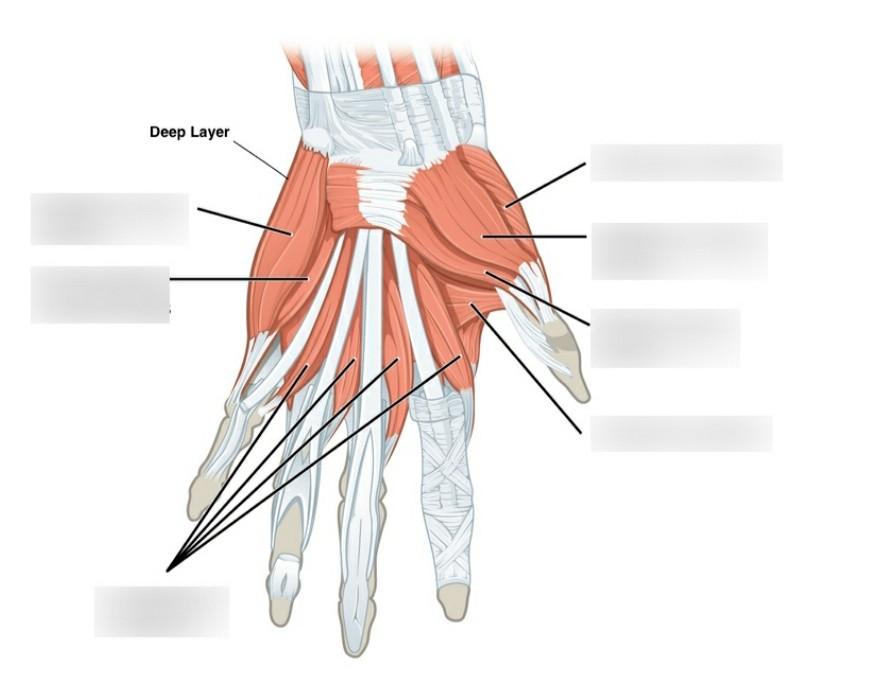
Effects Of Bedrest 5 The Muscles Joints And Mobility Nursing Times from cdn.ps.emap.com The extensor digitorum brevis muscle (sometimes edb) is a muscle on the upper surface of the foot that helps extend digits 2 through 4. Layer 3 of the foot. These are the tibialis anterior, extensor digitorum longus, extensor hallucis longus, and the peroneus tertius. • anterior compartment (innervated by the deep peroneal/anterior tibial nerve; The short and long muscles of the foot serve as synergists. Terms in this set (14). Neurovascular planes of the sole: The bones and joints in the feet experience wear and tear, so conditions that cause damage to the foot can directly affect its health.
Get 50% off osmosis prime.
Muscle layers of the sole of the foot. This article outlines the basic anatomy of the foot bones. The extensor digitorum brevis muscle (sometimes edb) is a muscle on the upper surface of the foot that helps extend digits 2 through 4. They are considered voluntary muscles. The foot is an intricate part of the body, consisting of 26 bones, 33 joints, 107 ligaments, and 19 muscles. Layer 3 of the foot. They are generally divided into two sets: These are the tibialis anterior, extensor digitorum longus, extensor hallucis longus, and the peroneus tertius. Try this amazing foot muscles anatomy: Those of the medial plantar region are connected with the great toe, and corrrespond with those of the thumb; The muscles are located mainly in the sole of the foot and divided into a central (medial) group and a group on either side (lateral). Sides of adjacent metatarsals i: Specialists in orthotics and foot scanning technology.
This means that the little toe can only be extended by the extensor digitorum longus muscle only. (a) the insertions of the flexor digitorum longus, flexor hallucis longus and little attention has been paid to the clinical assessment of intrinsic foot muscles in the musculoskeletal injury literature apart from few specific. These muscles insert on any one or several of the 26. These muscles begin and attach within the skeleton of the foot, have complex anatomical and topographical and functional relationships with the tendons of those leg muscles whose attachment points are on the bones of the foot. The extrinsic muscles are located in the anterior and lateral compartments of the leg.
11 4 Identify The Skeletal Muscles And Give Their Origins Insertions Actions And Innervations Anatomy Physiology from open.oregonstate.education Each foot has 26 bones that make up 33 joints and are held together by 19 muscles and 10 tendons and 107 ligaments! They retract the foot and effect. Neurovascular planes of the sole: Try this amazing foot muscles anatomy: Sides of adjacent metatarsals i: The dorsal aponeurosis of the toes supports the effect of the dorsal foot muscles by redirecting the force line of their tendons to. There are 19 or 20 intrinsic foot muscles, therefore 38 to 40 intrinsic foot muscle tendons. The bivalve foot, unlike that of gastropods, does not have a flat creeping sole but is bladelike (laterally the muscles mainly responsible for movement of the foot are the anterior and posterior pedal retractors.
Nearly a quarter of all bones in our bodies are in our feet.
The dorsal aponeurosis of the toes supports the effect of the dorsal foot muscles by redirecting the force line of their tendons to. (a) the insertions of the flexor digitorum longus, flexor hallucis longus and little attention has been paid to the clinical assessment of intrinsic foot muscles in the musculoskeletal injury literature apart from few specific. Get 50% off osmosis prime. The muscles at the top of the foot fan out to supply the individual toes. • anterior compartment (innervated by the deep peroneal/anterior tibial nerve; Flexor hallucis longus tendon transfer to the dorsum of the foot and release of the flexor digitorum longus and brevis tendons at the base of each toe. Calluses are created by friction applied to the skin of the at university foot and ankle institute, we take our patients' safety seriously. These include the three cuneiform bones, the cuboid bone, and the. There are 19 or 20 intrinsic foot muscles, therefore 38 to 40 intrinsic foot muscle tendons. They retract the foot and effect. Nearly a quarter of all bones in our bodies are in our feet. It connects the gastrocnemius and the soleus muscle with heel bone. Contrary to expectations, the intrinsic foot muscles contribute minimally to supporting the arch of the foot during walking and running.
Each foot has 26 bones that make up 33 joints and are held together by 19 muscles and 10 tendons and 107 ligaments! Insertions of the extrinsic foot muscle tendons on the plantar surface of the foot. These muscles begin and attach within the skeleton of the foot, have complex anatomical and topographical and functional relationships with the tendons of those leg muscles whose attachment points are on the bones of the foot. Foot muscle forces & deformities. To get started, all you need to do is click on the title of the article below that you are most interested in.
Solved Muscles Of The Lower Limb Foot 1 2 3 5 Dart Co Chegg Com from media.cheggcdn.com To get started, all you need to do is click on the title of the article below that you are most interested in. Their limited impact on posture and movement has led to the broad use of the extensor hallucis brevis and extensor digitorum brevis as muscular sources for tissue grafts. Muscle layers of the sole of the foot. Want a less stressful match week? Your foot and ankle specialist can shave down the thick layers of a callus using a scalpel blade. This is an online quiz called muscles of the foot. Muscles of the ankle and foot. However, these muscles do influence our ability to produce forward propulsion from one stride into the next, highlighting their role in bipedal locomotion.
The muscles acting on the foot can be divided into two distinct groups;
These muscles insert on any one or several of the 26. Like the muscles in the rest of the body, it's important to keep the muscles in the feet strong. Foot muscle forces & deformities. Terms in this set (14). They include the abductor halluces, the flexor digitorum brevis, the abductor digiti minimi, and the quadratus plantae. In the last post, we spoke of the ability of images to assist us as we learn complex tasks that involve the whole body. The muscles are located mainly in the sole of the foot and divided into a central (medial) group and a group on either side (lateral). Want a less stressful match week? Neurovascular planes of the sole: The muscles at the top of the foot fan out to supply the individual toes. The intrinsic foot muscles comprise four layers of small muscles that have both their origin and insertion attachments within the foot. Flexor hallucis longus tendon transfer to the dorsum of the foot and release of the flexor digitorum longus and brevis tendons at the base of each toe. Muscle layers of the sole of the foot.


0 Komentar